Author Nick Humphrey shares 5 key insights from his new book, Sentience: The Invention of Consciousness.
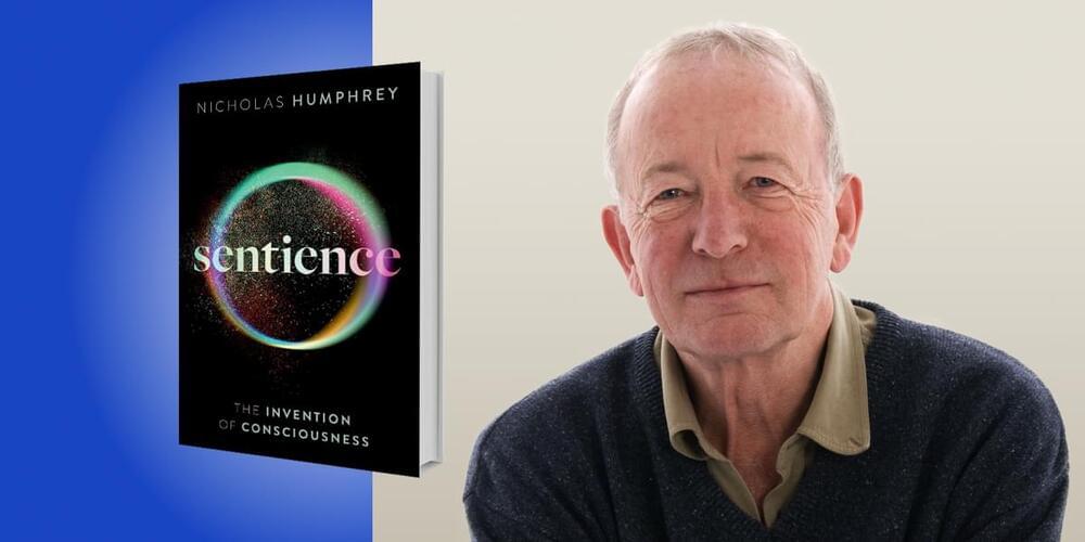
1. The mind, brain, and body are inextricably linked
The idea that the mind and brain are separate is usually attributed to the 17th-century French mathematician and philosopher René Descartes, who was what philosophers now call a substance dualist. Descartes believed that the mind and body are made of different substances: the body of a physical substance, and the mind of some mysterious, nonphysical material.
Today, most neuroscientists reject this idea. Modern brain research suggests that the mind is made of matter and emerges from brain activity. Even so, most still study the brain in isolation, without taking the body into consideration.
A wiring map diagrams more than half a million neuronal connections in the first complete connectome of Drosophila and holds clues about which brain architectures best support learning.

A small nucleus in the brainstem called locus coeruleus (literally the “blue spot,”) is the primary source of a major neuromodulator, norepinephrine (NE), an important mediator of the ‘fight or flight’ response in animals. However, very little is known about the local connections of this small albeit critically important group of neurons. A recent pioneering study published in eLife from the laboratory of Dr. Xiaolong Jiang, investigator at the Jan and Dan Duncan Neurological Research Institute (Duncan NRI) at Texas Children’s Hospital and assistant professor at Baylor College of Medicine, now reveals the cellular composition and circuit organization of the locus coeruleus in adult mice.
“In this study, we undertook the arduous task of mapping local connections of NE-producing neurons in the locus coeruleus,” Dr. Jiang said. “This is the first study of such an unprecedented magnitude and detail to be performed on the locus coeruleus, and in fact, on any monoamine neurotransmitter system. Our study has revealed that the neurons in the locus coeruleus have an unexpectedly rich cellular heterogeneity and local wiring logic.”
Locus coeruleus (LC) is known to house the vast majority of norepinephrine-releasing neurons in the brain and regulates many fundamental brain functions including the fight and flight response, sleep/wake cycles, and attention control. Present in the pontine region of the brainstem, LC neurons sense any existential dangers or threats in our external environment and send signals to alert other brain regions of the impending danger.
Breakthroughs don’t often happen in neuroscience, but we just had one. In a tour-de-force, an international team released the full brain connectivity map of the young fruit fly, described in a paper published last week in Science. Containing 3,016 neurons and 548,000 synapses, the map—called a connectome—is the most complex whole-brain wiring diagram to date.
“It’s a ‘wow,’” said Dr. Shinya Yamamoto at Baylor College of Medicine, who was not involved in the work.
Why care about a fruit fly? Far from uninvited guests at the dinner table, Drosophila melanogaster is a neuroscience darling. Although its brain is smaller than a poppy seed—a far cry from the 100 billion neurons that power human brains—the fly’s neural system shares similar principles to those that underlie our own brains.

Two new effects on TikTok can give users sculpted cheekbones, plumped lips, or a younger look with the push of a button. But this hyper-realistic image-altering tech also spurs backlash. WSJ reporter Sara Ashley O’Brien joins host Zoe Thomas to discuss how these filters work and why some experts say they could damage users’ mental health. Photo: Storyblocks.
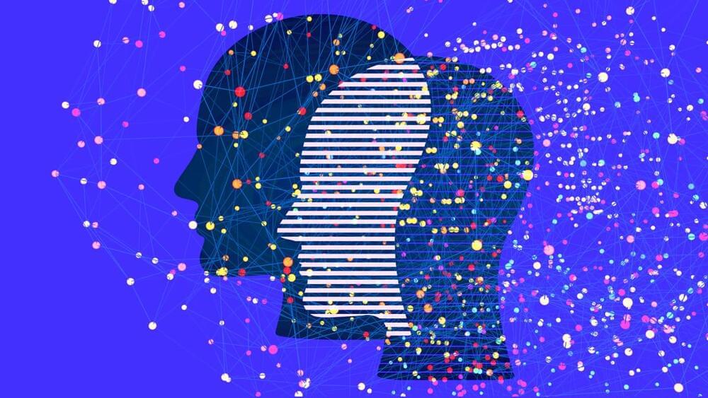
This video explores what attention really is, what role it plays in learning and why people can’t multitask — the issue of attention residue.
OUTLINE:
0:00 — Sneak peek.
0:20 — Introduction.
0:57 — Why we need attention.
1:46 — Thalamus as attentional filter.
3:06 — Higher attentional systems.
3:40 — Role of attention in learning.
4:42 — Attention residue.
6:00 — Conclusions and references.
Socials:
VK: https://vk.com/atpsynthase