Quantum computers get a lot of attention, even though they are not ready for prime time, but quantum sensors are already doing useful work. These sensors measure fields, forces and motion so small that ordinary background noise can drown them out. Some sensors are already in daily use, while others are moving from research labs into flight tests, hospitals and field instruments.
For example, a human brain produces magnetic signals in the femtotesla-to-picotesla range—billions of times weaker than a refrigerator magnet—far weaker than the magnetic noise in an ordinary room. That is why brain scanners that measure these signals need ultrasensitive detectors and strong magnetic shielding. In some hospitals, these detectors use quantum technology to help map brain activity before epilepsy surgery, without touching the brain.
Quantum sensors are showing up in other fields as well, including in navigation when GPS signals are jammed or spoofed, mapping gravity to reveal what’s underground, and boosting astronomers’ ability to measure gravitational waves. I am a photonics and quantum technologies researcher. My lab applies physics to develop a range of devices, including quantum sensors.
Lisa Feldman Barrett, Michael Levin and Adam Frank discuss whether science should abandon its materialist framework.
Could a different metaphysics help science to progress further?
With a free trial, you can watch the full debate NOW at https://iai.tv/video/science-beyond-t… centuries, we’ve assumed that science has banished the transcendent and established that reality is entirely physical. But critics argue there are signs that a rigorous materialism might be holding science back. Increasingly, “emergence” is used to account for everything from consciousness to spacetime – a convenient placeholder for what materialist science may be unable to explain. Physicists like Heisenberg and Hawking concluded that science gives us models of reality, rather than final descriptions of its true nature, while there are scientists working in everything from biology to computer science who suggest that dualism is a productive metaphysical framework for their research. Materialism may have enabled science to reach beyond the dogmas of religion, but there are now those who are restlessly probing the limits of materialism itself. Does science need to assume a materialist account of the world or might this have fundamental limitations? Could a different metaphysics help science make progress on key questions, from the origin of life to the mysteries of quantum gravity? Or would abandoning materialism risk returning us to the myths of superstition and religion? #science #materialism #metaphysics Lisa Feldman Barrett is among the most cited scientists in the world for her research on the psychology and neuroscience of emotions. Adam Frank is an astrophysicist who explores the origins of stars, civilizations and consciousness, and is a leading figure in astrobiology and the search for alien life. Michael Levin is a synthetic biologist whose pioneering work in regenerative biology involves building biological robots to probe the nature of life, intelligence and evolution. Güneş Taylor hosts. The Institute of Art and Ideas features videos and articles from cutting edge thinkers discussing the ideas that are shaping the world, from metaphysics to string theory, technology to democracy, aesthetics to genetics. Subscribe today! https://iai.tv/subscribe?utm_source=Y… 0:00 Intro 1:34 Science cannot reveal objective reality 5:28 — History shows that materialism is one of many philosophies of science 8:59 There are some mathematical facts which are discovered, not chosen 12:14 Does materialism prevent mythical and superstitious views of reality? 14:56 There is no 3rd person view of the universe 18:05 Is science truly reproducible? For debates and talks: https://iai.tv For articles: https://iai.tv/articles For courses: https://iai.tv/iai-academy/courses.
For centuries, we’ve assumed that science has banished the transcendent and established that reality is entirely physical. But critics argue there are signs that a rigorous materialism might be holding science back. Increasingly, “emergence” is used to account for everything from consciousness to spacetime – a convenient placeholder for what materialist science may be unable to explain. Physicists like Heisenberg and Hawking concluded that science gives us models of reality, rather than final descriptions of its true nature, while there are scientists working in everything from biology to computer science who suggest that dualism is a productive metaphysical framework for their research. Materialism may have enabled science to reach beyond the dogmas of religion, but there are now those who are restlessly probing the limits of materialism itself.
Does science need to assume a materialist account of the world or might this have fundamental limitations? Could a different metaphysics help science make progress on key questions, from the origin of life to the mysteries of quantum gravity? Or would abandoning materialism risk returning us to the myths of superstition and religion?
Aging human breast atlas reveals cancer susceptibility
The team used advanced imagining techniques to analyse breast tissue from more than 500 women aged 15 to 86 years old. The tissue included biopsies taken from women for non-cancer-related reasons.
Combining these images with details of the hormone receptors and immune cells present, as well as the tissue architecture, the researchers were able to map how breast tissue changes over time in unprecedented detail. Their findings point to reasons why breast cancer risk increases with age and why tumors in younger women differ biologically.
The author added: “Our map revealed that as women age, their breast tissue goes through major changes, with the most dramatic changes occurring at menopause. There are changes, too, during their twenties, possibly linked to pregnancy and childbirth, but these are far less pronounced.”
The map revealed that all types of cells become fewer in number and divide far less often. Milk-producing structures known as lobules shrink or disappear, while the ducts that that carry milk become relatively more common, with the supporting layer around them becoming thicker. Fat cells increase while blood vessels decrease.
Meanwhile, changes occur in the immune environment. Younger breasts have more B cells and active T cells, which helps them identify and kill cancer cells. As tissue ages, these types of cells decline in number, replaced by other types of immune cell that indicate a more inflammatory and potentially less protective immune environment. ScienceMission sciencenewshighlights.
Scientists at the UCLA Health Jonsson Comprehensive Cancer Center have developed a new cytokine-armored CAR-T cell therapy that helps the immune system better attack aggressive brain tumors in mice while reducing dangerous side effects that have long limited immune-based treatments for glioblastoma, one of the deadliest and most treatment-resistant brain cancers.
The therapy works by reprogramming CAR-T cells to release immune-stimulating proteins, called IL-12 and DR-18, that activate the body’s own immune system, strengthening the overall anti-cancer response. In mouse models, the approach improved tumor control, including against cancers made up of mixed cell populations that often escape therapies.
Researchers also found that pairing the treatment with a second CAR-T strategy targeting VEGF, a protein that drives abnormal blood vessel growth and contributes to swelling in glioblastoma, helped reduce side effects while preserving strong anti-tumor activity.
Scientists may have found the brain’s “pain switch”—and how to turn it off. New research from the University of Colorado Boulder points to a little-known brain circuit that may determine whether short-term pain fades away or becomes a long-lasting problem. The findings suggest that this pathway plays a key role in turning temporary pain into chronic pain that can persist for months or even years.
The study, conducted in animals and published in the Journal of Neuroscience, focused on a region called the caudal granular insular cortex (CGIC). Researchers found that shutting down this circuit can both prevent chronic pain from developing and stop it after it has already begun.
“Our paper used a variety of state-of-the art methods to define the specific brain circuit crucial for deciding for pain to become chronic and telling the spinal cord to carry out this instruction,” said senior author Linda Watkins, distinguished professor of behavioral neuroscience in the College of Arts and Sciences. “If this crucial decision maker is silenced, chronic pain does not occur. If it is already ongoing, chronic pain melts away.”
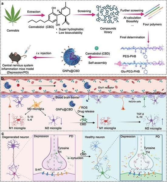
Breakthrough in brain medicine: a new way to deliver CBD!
Cannabidiol (CBD) has incredible potential to fight brain inflammation, but it has always faced a major roadblock: it struggles to dissolve and cross the blood-brain barrier. Researchers have just developed an ingenious solution using glucose-coated nanoparticles to get CBD exactly where it needs to go.
Here’s why it’s a game-changer: 🔬 Sneaky Delivery: The glucose coating helps the particles “hitch a ride” on the brain’s natural glucose transporters, successfully smuggling the CBD across the blood-brain barrier. 🎯 Smart Release: Once inside the brain, the nanoparticles target immune cells (microglia) and only release the CBD when they detect the chemical stress of active inflammation. 🐁 Promising Results: In mouse models of Parkinson’s disease and depression, this new delivery method drastically reduced inflammation, protected neurons, and improved behavioral recovery compared to standard CBD.
This targeted approach could be a massive step forward in treating chronic neuroinflammatory diseases! 🧬✨
Studty.
Glucose-coated nanoparticles carry CBD across the blood-brain barrier, trigger release in inflamed tissue, and reduce neuroinflammatory signs in mice.
Roles of lysosomal small-molecule transporters in metabolism and signaling
Small-molecule transporters of the lysosomal membrane export lysosomal catabolites for reuse in cell metabolism.
These transporters often show substrate promiscuity and, conversely, a given metabolite is often exported through distinct transport routes and sometimes in different states (e.g., single amino acids versus dipeptides).
Some lysosomal transporters import metabolites into the lumen. The combination of importers and exporters can create small-molecule shuttles across the lysosomal membrane, which regulate the lumen state.
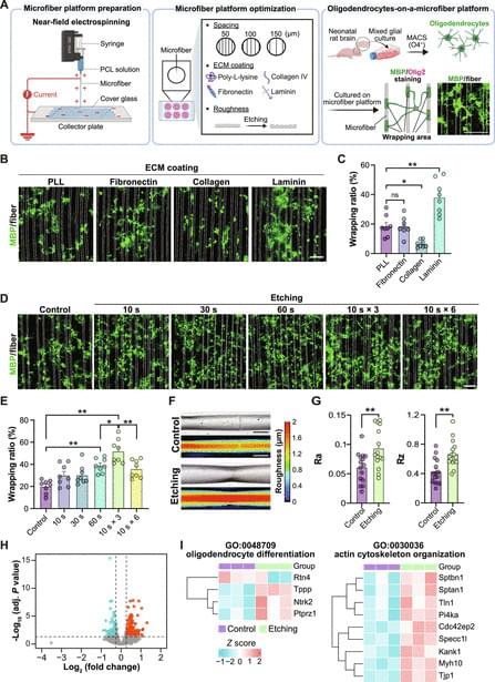
Remyelination requires the precise wrapping of axons by oligodendrocyte processes, a critical step for restoring neural circuit function. However, a lack of quantitative systems that recapitulate axonal geometry and chemistry has limited mechanistic and pharmacological insights into myelin wrapping. Here, we present a bioengineered microfiber platform that mimics neurite architecture and surface chemistry, enabling high-content quantification of oligodendrocyte wrapping. Through compound screening, we identified dimemorfan, a clinically used sigma-1 receptor agonist, as a potent enhancer of myelin wrapping. Dimemorfan treatment accelerated remyelination and functional recovery in demyelinated mice and promoted myelin wrapping by human induced pluripotent stem cell (iPSC)-derived oligodendrocytes.
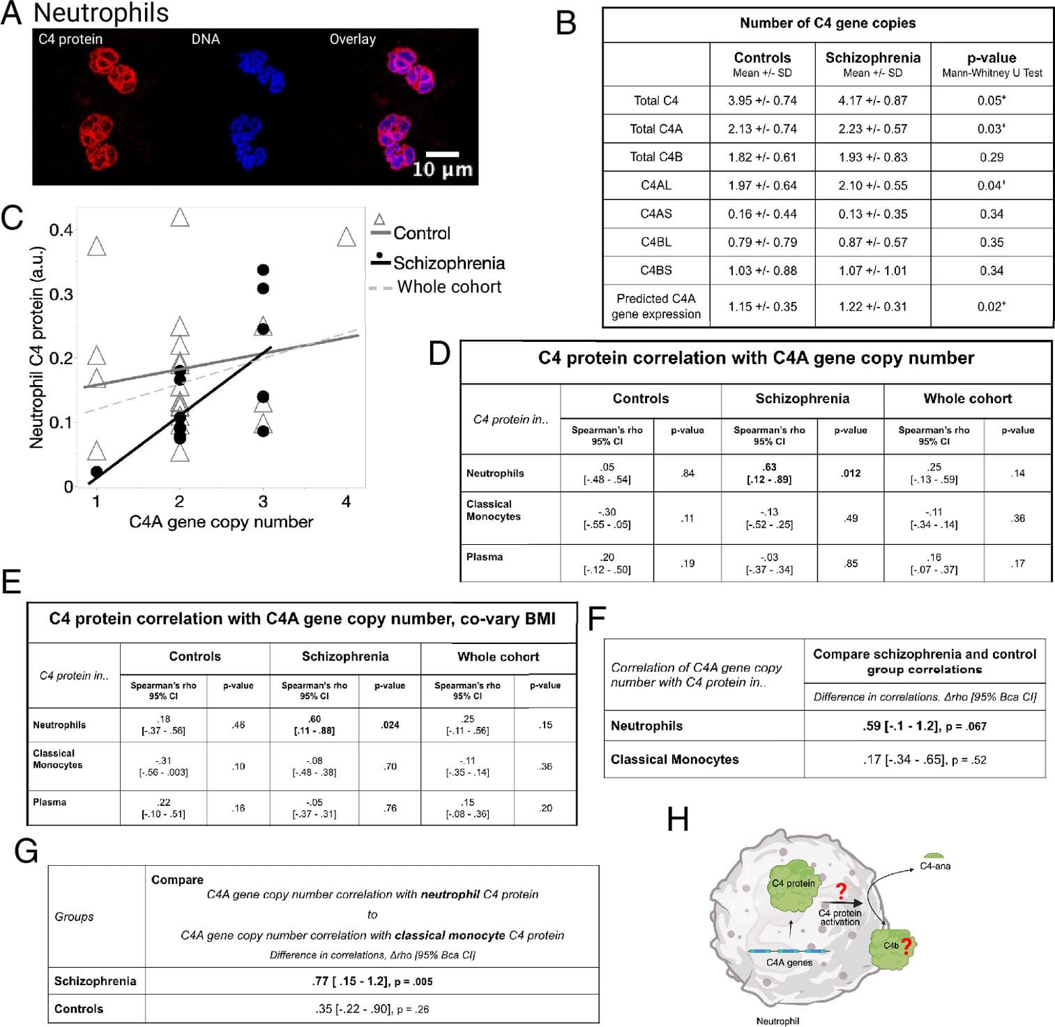
The most common white blood cells in your body—immune cells called neutrophils—can make a protein nobody knew they were making, Stanford Medicine investigators have discovered. That unexpected sighting joins a growing list of hints tying schizophrenia, a disorder of the brain, to events occurring elsewhere in our bodies. The findings are summarized in a paper published in Proceedings of the National Academy of Science.
The newly noticed neutrophil nexus, as a source of the protein called C4A, links a long list of other observations that are somewhat puzzling when looked at in isolation: For example, large-scale population-genetic studies have identified C4A, already known to be produced mainly in the liver, as a pronounced risk factor in schizophrenia. People with schizophrenia tend to have increased numbers of neutrophils in their blood. And the most effective medication for schizophrenia inhibits neutrophils.
Schizophrenia affects one in every 100 persons globally almost without variation by geography or ethnicity. Its most noticeable symptoms are hallucinations, delusions and fixations. A fundamental feature of the disease is cognitive impairment: inability to think clearly, reduced working memory, disorganized thinking and behavior.