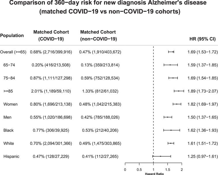
Toggle light / dark theme

Human Induced Pluripotent Stem Cells : Clinical Significance and Applications in Neurologic Diseases

face_with_colon_three could heal body parts in humans.


The generation of human induced pluripotent stem cells (iPSCs) from somatic cells using gene transfer opens new areas for precision medicine with personalized cell therapy and encourages the discovery of essential platforms for targeted drug development. iPSCs retain the genome of the donor, may regenerate indefinitely, and undergo differentiation into virtually any cell type of interest using a range of published protocols. There has been enormous interest among researchers regarding the application of iPSC technology to regenerative medicine and human disease modeling, in particular, modeling of neurologic diseases using patient-specific iPSCs. For instance, Parkinson’s disease, Alzheimer’s disease, and spinal cord injuries may be treated with iPSC therapy or replacement tissues obtained from iPSCs. In this review, we discuss the work so far on generation and characterization of iPSCs and focus on recent advances in the use of human iPSCs in clinical setting.

Stem cells exhibit the capacity of self-renewal and may undergo differentiation into various tissue types. These are divided into pluripotent stem cells (PSCs; embryonic stem cells [ESCs] and induced pluripotent stem cells [iPSCs]) and multipotent stem cells (adult stem cells [ASCs]) based on their differentiation capacity [45]. PSCs, including ESCs derived from embryos and iPSCs derived by gene transfer, may undergo indefinite proliferation and differentiate into different types of tissues depending on the treatment conditions [86]. Multipotent stem cells, however, may be obtained from tissue-derived precursors (umbilical cord blood, bone marrow, adipose tissue, placenta, or blood), which are already grown tissues.

Advance of Stem Cell Treatment for Traumatic Brain Injury

Traumatic brain injury (TBI) is an important cause of human mortality and morbidity, which can induce serious neurological damage. At present, clinical treatments for neurological dysfunction after TBI include hyperbaric oxygen, brain stimulation and behavioral therapy, but the therapeutic effect is not satisfactory. Recent studies have found that exogenous stem cells can migrate to damaged brain tissue, then participate in the repair of damaged brain tissue by further differentiation to replace damaged cells, while releasing anti-inflammatory factors and growth factors, thereby significantly improving neurological function. This article will mainly review the effects, deficiencies and related mechanisms of different types of stem cells in TBI.

Traumatic brain injury (TBI) is a common and frequently occurring disease. According to the World Health Organization, TBI will become the main cause of human mortality and morbidity after 2020, which brings a heavy economic burden to patients and families (Maas et al., 2017). TBI is a disease which causes the destruction of normal brain function, and leads to serious physical, cognitive and emotional disorders. The pathophysiology of TBI mainly includes the break of the blood brain barrier (BBB), extensive neuroinflammation, diffuse axonal injury, and neurodegenerative lesions (Xiong et al., 2008). The pathological changes of brain injury are mainly the loss of normal tissue structure, destruction of neuronal cells and internal environment disturbance, among which neuronal cells injury is the key point. There is no effective drug treatment so far.

How does the brain link events to form a memory? Study reveals unexpected mental processes

A woman walking down the street hears a bang. Several moments later she discovers her boyfriend, who had been walking ahead of her, has been shot. A month later, the woman checks into the emergency room. The noises made by garbage trucks, she says, are causing panic attacks. Her brain had formed a deep, lasting connection between loud sounds and the devastating sight she witnessed.

This story, relayed by clinical psychiatrist and co-author of a new study Mohsin Ahmed, MD, Ph.D., is a powerful example of the ’s powerful ability to remember and connect events separated in time. And now, in that new study in mice published today in Neuron, scientists at Columbia’s Zuckerman Institute have shed light on how the brain can form such enduring links.

The scientists uncovered a surprising mechanism by which the hippocampus, a brain region critical for memory, builds bridges across time: by firing off bursts of activity that seem random, but in fact make up a complex pattern that, over time, help the brain learn associations. By revealing the underlying circuitry behind associative learning, the findings lay the foundation for a better understanding of anxiety and trauma- and stressor-related disorders, such as panic and post-traumatic stress disorders, in which a seemingly neutral event can elicit a negative response.

Heart–brain interactions shape somatosensory perception and evoked potentials

Our brain continuously receives signals from the body and the environment. Although we are mostly unaware of internal bodily processes, such as our heartbeats, they can affect our perception. Here, we show two distinct ways in which the heartbeat modulates conscious perception. First, increased heartbeat-evoked neural activity before stimulation is followed by decreased somatosensory detection. This effect can be explained by subjects adopting a more conservative decision criterion, which is accompanied by changes in early and late somatosensory-evoked responses. Second, stimulus timing during the cardiac cycle affects sensitivity but not criterion for somatosensory stimuli, which is reflected only in late somatosensory-evoked responses. We propose that these heartbeat-related modulations are connected to fluctuations of interoceptive attention and (unconscious) predictive coding mechanisms.

Even though humans are mostly not aware of their heartbeats, several heartbeat-related effects have been reported to influence conscious perception. It is not clear whether these effects are distinct or related phenomena, or whether they are early sensory effects or late decisional processes. Combining electroencephalography and electrocardiography, along with signal detection theory analyses, we identify two distinct heartbeat-related influences on conscious perception differentially related to early vs. late somatosensory processing. First, an effect on early sensory processing was found for the heartbeat-evoked potential (HEP), a marker of cardiac interoception. The amplitude of the prestimulus HEP negatively correlated with localization and detection of somatosensory stimuli, reflecting a more conservative detection bias (criterion).

A new high-resolution, 3D map of the whole mouse brain

After three years of intensive data-gathering and careful drawing, the mapmakers’ work was complete.

The complex terrain they charted, with all its peaks, valleys and borders, is only about half an inch long and weighs less than a jellybean: the brain of the laboratory mouse.

In a paper published today in the journal Cell, the Allen Institute mapmakers describe this cartographical feat—the third iteration of the Allen Mouse Brain Common Coordinate Framework, or CCFv3 (https://portal.brain-map.org/), a complete, high-resolution 3D atlas of the mouse brain.