Researchers from Brigham and Women’s Hospital have engineered yeast used in baking, wine-making and brewing to treat inflammatory bowel disease (IBD). The bacteria has been modified to secrete an anti-inflammatory molecule in response to signs of gut inflammation and has proven effective in preclinical tests.
Our gut microbiome is increasingly implicated in everything from cancer to neurodegenerative disease but it is still unclear exactly how we can translate these novel findings into clinical treatments. Fecal transplants are probably the most primitive microbiome-modifying treatment we have developed, while probiotics simply rely on upping specific levels of naturally occurring bacteria.
Perhaps the most futurist microbiome therapy under investigation is the idea of genetically engineered probiotics. Here researchers modify bacteria to either eat up molecules we don’t want in our body or secrete molecules we know have positive therapeutic effects.
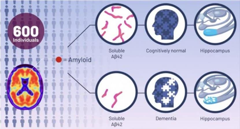
Summary: Study finds amyloid-beta plaques may not be the cause of memory loss associated with Alzheimer’s disease, but instead a consequence of the disease. Regardless of the levels of amyloid plaques, researchers found individuals with high levels of amyloid peptide were cognitively normal. Higher levels of soluble amyloid beta peptide were also linked to people having a larger hippocampus.
Source: University of Cincinnati.
Experts estimate more than 6 million Americans are living with Alzheimer’s dementia. But a recent study, led by the University of Cincinnati, sheds new light on the disease and a highly debated new drug therapy.
Scientists develop the first CRISPR-Cas9-based gene drive in plants which may breed crops better able to withstand drought and disease.
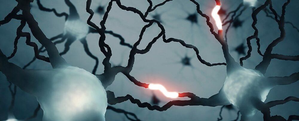
Scientists have discovered a unique form of cell messaging occurring in the human brain that’s not been seen before. Excitingly, the discovery hints that our brains might be even more powerful units of computation than we realized.
Early last year, researchers from institutes in Germany and Greece reported a mechanism in the brain’s outer cortical cells that produces a novel ‘graded’ signal all on its own, one that could provide individual neurons with another way to carry out their logical functions.
By measuring the electrical activity in sections of tissue removed during surgery on epileptic patients and analysing their structure using fluorescent microscopy, the neurologists found individual cells in the cortex used not just the usual sodium ions to ‘fire’, but calcium as well.
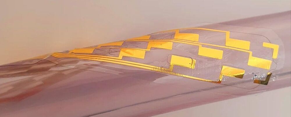
Scientists have revealed a fascinating new design for an incredibly tiny, inflatable spinal cord implant, suited for treating severe chronic back pain that doesn’t respond to medication.
The inflatable electronic device is part of a spinal cord stimulator (SCS) setup, a type of well-established therapy that delivers mild electric currents to a person’s spinal cord via implanted electrodes. That current is sent by a small, implanted pulse generator device, and the whole thing reduces pain because the electrical pulses help to mask pain signals traveling to the brain via the spinal cord.
If that all sounds rather invasive, that’s because it is. But this new device, designed by a team led by scientists from the University of Cambridge in the UK, could help to change that — with less invasive surgery requirements.
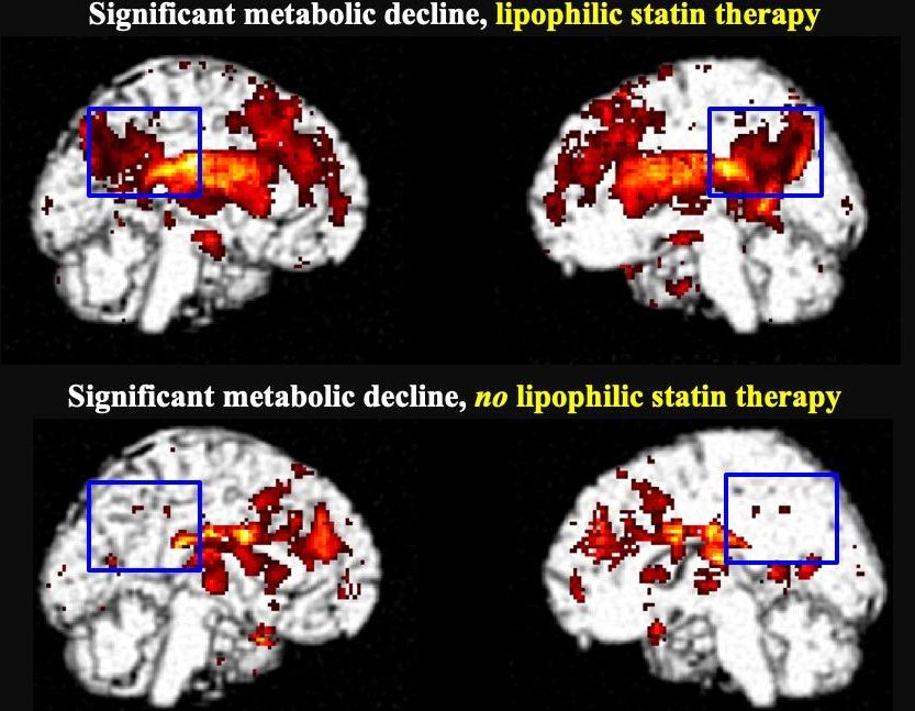
In patients with mild cognitive impairment, taking lipophilic statins more than doubles their risk of developing dementia compared to those who do not take statins. According to research presented at the Society of Nuclear Medicine and Molecular Imaging 2021 Annual Meeting, positron emission tomography (PET) scans of lipophilic statin users revealed a highly significant decline in metabolism in the area of the brain that is first impacted by Alzheimer’s disease.
Statins are medications used to lower cholesterol and reduce the risk of heart attack or stroke. They are the most commonly used drugs in the developed world, and nearly 50 percent of Americans over age 75 use a statin. Different types of statins are available based on a patient’s health needs, including hydrophilic statins that focus on the liver and lipophilic statins that are distributed to tissues throughout the body.
Lipophilic statins include simvastatin, fluvastatin, pitavastatin, lovastatin and atorvastatin. Hydrophilic statins include rosuvastatin and pravastatin.
Flexible thinking is key to creativity – in other words, the ability to think of new ideas, make novel connections between ideas, and make new inventions. It also supports academic and work skills such as problem solving. That said, unlike working memory – how much you can remember at a certain time – it is largely independent of IQ, or “crystallised intelligence”.
IQ is often hailed as a crucial driver of success, particularly in fields such as science, innovation and technology. In fact, many people have an endless fascination with the IQ scores of famous people. But the truth is that some of the greatest achievements by our species have primarily relied on qualities such as creativity, imagination, curiosity and empathy.
Many of these traits are embedded in what scientists call “cognitive flexibility” – a skill that enables us to switch between different concepts, or to adapt behaviour to achieve goals in a novel or changing environment. It is essentially about learning to learn and being able to be flexible about the way you learn. This includes changing strategies for optimal decision-making. In our ongoing research, we are trying to work out how people can best boost their cognitive flexibility.
Cognitive flexibility provides us with the ability to see that what we are doing is not leading to success and to make the appropriate changes to achieve it. If you normally take the same route to work, but there are now roadworks on your usual route, what do you do? Some people remain rigid and stick to the original plan, despite the delay. More flexible people adapt to the unexpected event and problem-solve to find a solution.
CRISPR gene editing already promises to fight diseases that were once thought unassailable, but techniques so far have required injecting the tools directly into affected cells. That’s not very practical for some conditions. However, there’s just been a breakthrough. NPRreports that researchers have published results showing that you can inject CRISPR-Cas9 into the bloodstream to make edits, opening the door to the use of gene editing for treating many common diseases.
The experimental treatment tackled a rare genetic disease, transthyretin amyloidosis. Scientists injected volunteers with CRISPR-loaded nanoparticles that were absorbed by the patients’ livers, editing a gene in the organ to disable production of a harmful protein. Levels of that protein plunged within weeks of the injection, saving patients from an illness that can rapidly destroy nerves and other tissues in their bodies.
The test involved just six people, and the research team still has to conduct long-term studies to check for possible negative effects. If this method proves viable on a large scale, though, it could be used to treat illnesses where existing CRISPR techniques aren’t practical, ranging from Alzheimer’s to heart disease.
Neil deGrasse Tyson explains the early state of our Universe. At the beginning of the universe, ordinary space and time developed out of a primeval state, where all matter and energy of the entire visible universe was contained in a hot, dense point called a gravitational singularity. A billionth the size of a nuclear particle.
While we can not imagine the entirety of the visible universe being a billion times smaller than a nuclear particle, that shouldn’t deter us from wondering about the early state of our universe. However, dealing with such extreme scales is immensely counter-intuitive and our evolved brains and senses have no capacity to grasp the depths of reality in the beginning of cosmic time. Therefore, scientists develop mathematical frameworks to describe the early universe.
Neil deGrasse Tyson also mentions that our senses are not necessarily the best tools to use in science when uncovering the mysteries of the Universe.
It is interesting to note that in the early Universe, high densities and heterogeneous conditions could have led sufficiently dense regions to undergo gravitational collapse, forming black holes. These types of Primordial black holes are hypothesized to have formed soon after the Big Bang. Going from one mystery to the next, some evidence suggests a possible Link Between Primordial Black Holes and Dark Matter.
In modern physics, antimatter is made up of elementary particles, each of which has the same mass as their corresponding matter counterparts — protons, neutrons and electrons — but the opposite charges and magnetic properties.
A collision between any particle and its anti-particle partner leads to their mutual annihilation, giving rise to various proportions of intense photons, gamma rays and neutrinos. The majority of the total energy of annihilation emerges in the form of ionizing radiation. If surrounding matter is present, the energy content of this radiation will be absorbed and converted into other forms of energy, such as heat or light. The amount of energy released is usually proportional to the total mass of the collided matter and antimatter, in accordance with Einstein’s mass–energy equivalence equation.
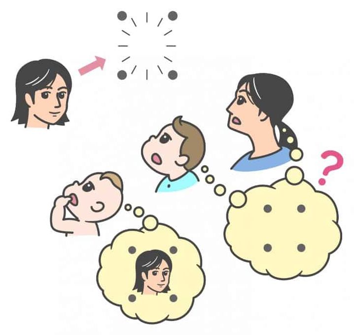
The results of the study demonstrated that the mechanisms for visual perception change drastically in the second half of the first year of life, from the bottom-up system to the system incorporating top-down processing.
Summary: Study reveals very young infants can perceive objects that older infants, children, and adults can not see due to a phenomenon called visual backward masking.
Source: Chuo University
We can generally recognize an object, even if it is presented for a very brief time. However, if another object appears immediately following the first object, the perception on the first object is impaired such that we do not notice its existence.
This perceptual phenomenon, called “visual backward masking,” is used in vision science to study how visual perception is processed in the brain. Interestingly, this phenomenon occurs even if the second object does not spatially overlap the first object, such as a contour or four dots surrounding the object.
STANFORD, Calif. — A groundbreaking “superhero” vaccine inspired by the DNA code of Olympic athletes could help transform society over the next decade, a top genetic scientist claims.
The vaccine would provide lifelong protection against three of the top ten leading causes of death, according to Euan Ashley, professor of medicine and genetics at Stanford University. The so-called “superhero” jab could offer simultaneous, long-term protection against heart disease, stroke, Alzheimer’s disease, and liver disease, thanks to advances in genetic engineering.
This breakthrough treatment would deliver the blueprint of “ideal” cells from men and women whose genes are more disease-resistant than those of the average person, together with an “instruction manual” to help the body “repair, tweak and improve” its own versions. A single dose could lead to a “body-wide genetic upgrade” that would cut the risk of premature death in some adults by as much as 50 percent.