Toggle light / dark theme

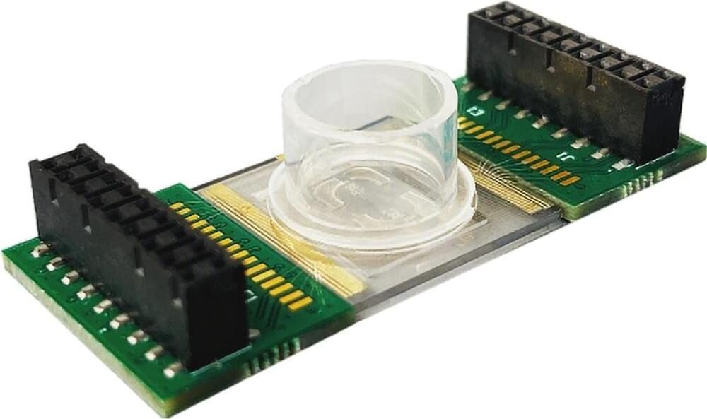
Researchers at the University of Massachusetts Amherst have developed an innovative technology inspired by the synchronization mechanism of WWI fighter aircraft, which coordinated machine gun fire with propeller movement. This breakthrough allows precise, real-time control of the pH in a cell’s environment to influence its behavior. Detailed in Nano Letters, the study opens exciting possibilities for developing new cancer and heart disease therapies and advancing the field of tissue engineering.

“Every cell is responsive to pH,” explains Jinglei Ping, associate professor of mechanical and industrial engineering at UMass Amherst and corresponding author of the study. “The behavior and functions of cells are impacted heavily by pH. Some cells lose viability when the pH has a certain level and for some cells, the pH can change their physiological properties.” Previous work has demonstrated that changes of pH as small as 0.1 pH units can have physiologically significant effects on cells.
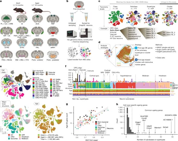
Sensitive cells: Scientists discovered dozens of specific cell types, mostly glial cells, known as brain support cells, that underwent significant gene expression changes with age. Those strongly affected included microglia and border-associated macrophages, oligodendrocytes, tanycytes, and ependymal cells.

Inflammation and neuron protection: In aging brains, genes associated with inflammation increased in activity while those related to neuronal structure and function decreased.

Aging hot spot: Scientists discovered a specific hot spot combining both the decrease in neuronal function and the increase in inflammation in the hypothalamus. The most significant gene expression changes were found in cell types near the third ventricle of the hypothalamus, including tanycytes, ependymal cells, and neurons known for their role in food intake, energy homeostasis, metabolism, and how our bodies use nutrients. This points to a possible connection between diet, lifestyle factors, brain aging, and changes that can influence our susceptibility to age-related brain disorders.

Brain-wide cell-type-specific transcriptomic signatures of healthy ageing in mice.


We termed enhancers that gained (and maintained) H3K4me1 in obesity and WL ‘new enhancers’. Most of these ‘new enhancers’ were also active (that is, marked by H3K27ac) during obesity and/or WL (Fig. 4D). We then annotated the enhancers to their closest gene and performed a GSEA. In agreement with the promoter GSEA above, we found that the ‘new active enhancers’ were related to inflammatory signalling, lysosome activity and extracellular matrix remodelling (Fig. 4e and Extended Data Fig. 9i), indicating a persistent shift of adipocytes towards a more inflammatory and less adipogenic identity. Corroborating these results, Roh et al. had analysed H3K27ac in adipocytes of obese mice and reported impaired identity maintenance during obesity25.

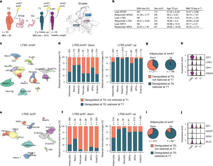
To combine our findings regarding retained translational changes and epigenetic memory, we investigated whether epigenetic mechanisms, such as differentially marked promoters or enhancers, could explain the persistent translational obesity-associated changes after WL. Notably, 57–62% of downregulated and 68–75% of upregulated persistent translational DEGs after WL could be accounted for by one or more of the analysed epigenetic modalities (Fig. 4f). Overall, these results strongly suggest the presence of stable cellular, epigenetic and transcriptional memory in mouse adipocytes that persists after WL.

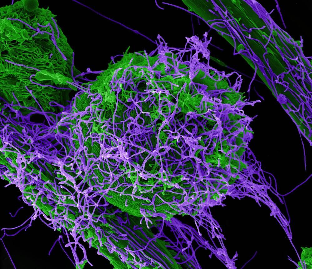
Ebola is a deadly hemorrhagic disease caused by a virus that is endemic in parts of East-Central and West Africa. Most people are aware that a primary route for person-to-person transmission is through contact with bodily fluids from an infected person. But more recent outbreaks, including the 2013–2016 Ebola epidemic in West Africa, demonstrated that infectious Ebola virus (EBOV) is also found on the skin’s surface of those who have succumbed to infection or at late times during infection.

Although evidence suggests that EBOV can be passed on from skin contact with a person in the later stages of the disease, very little is known about how the virus makes its way out of the body and onto the skin’s surface.

Researchers at University of Iowa Health Care and colleagues at Texas Biomedical Research Institute and Boston University have traced a cellular route the virus uses to traverse the inner and outer layers of skin and emerge onto the skin’s surface.

Researchers have developed a method to direct stem cells to form specific structures. By triggering the expression of specific genes in mouse embryonic stem cells, synthetic organizer cells were created, which can assemble in specific ways and carry out various phsyiological functions. This work is an important step on the road to eventually using synthetic cells to repair damaged tissues or regenerate organs. The research has been reported in Cell.

The researchers created synthetic organizer cells that could generate a structure like a mouse body, from head to tail, that underwent processes that were similar to those in mouse embryonic development. Another type of synthetic organizer cell was used to produce a structure that was similar to a heart, and featured a central chamber. This synthetic, heart-like structure also had a network of blood vessels and beat regularly.

New research identifies E-TCmito as a key link between neuronal activity and mitochondrial function, highlighting its potential to address cognitive decline in aging and diseases like Alzheimer’s.

New research in mice has identified a critical mechanism that connects neuronal activity with mitochondrial function, offering insight into potential strategies to address age-related cognitive decline. Mitochondria, essential for meeting the energy needs of active neurons, generate adenosine triphosphate (ATP) primarily through oxidative phosphorylation (OXPHOS).

As mammals age, the efficiency of mitochondrial metabolism in the brain declines, significantly impacting neuronal and network function. The disruption of the OXPHOS pathway contributes to oxidative stress and mitochondrial dysfunction, exacerbating these challenges.

Donald J. Cram, a Nobel Prize-winning chemist who taught andconducted research at UCLA for more than 50 years and is remembered bythousands of undergraduates for singing and playing guitar in class, died ofcancer June 17 at his home in Palm Desert. He was 82.

A renowned scientist who was as comfortable riding the waveswith friends in the San Onofre Surfing Club as he was in his lab at UCLAconstructing complex molecular models, Cram won the Nobel Prize in 1987 and theNational Medal of Science in 1993 for his work in host-guest chemistry, a fieldhe helped to create. In 1998, he wasranked among the 75 most important chemists of the past 75 years byChemical and Engineering News.

“DonaldCram stands alone in the incredible variety, beauty and depth of hisaccomplishments,” read the citation for Cram’s National Medal of Science. “His investigations have helped give thisscience its form and sophistication. Hetruly brought art to science by making his science an art.”

Penn Engineers have modified lipid nanoparticles (LNPs) — the revolutionary technology behind the COVID-19 mRNA vaccines — to not only cross the blood-brain barrier (BBB) but also to target specific types of cells, including neurons. This breakthrough marks a significant step toward potential next-generation treatments for neurological diseases like Alzheimer’s and Parkinson’s.

In a new paper in Nano Letters, the researchers demonstrate how peptides — short strings of amino acids — can serve as precise targeting molecules, enabling LNPs to deliver mRNA specifically to the endothelial cells that line the blood vessels of the brain, as well as neurons.

This represents an important advance in delivering mRNA to the cell types that would be key in treating neurodegenerative diseases; any such treatments will need to ensure that mRNA arrives at the correct location. Previous work by the same researchers proved that LNPs can cross the BBB and deliver mRNA to the brain, but did not attempt to control which cells the LNPs targeted.